
Ultrasound case library
Real, anonymized and captioned cases from daily medical practice. Two cases are fully open, with high-definition images and videos; for the others, the text and thumbnail are public and HD media opens once you sign in.
Why this archive matters
This is not an internet image bank. It is a curated slice of real clinical routine with captions, context and ultrasound vocabulary.
 HD for signed-in usersÚtero
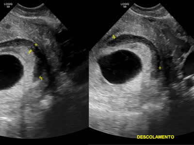
Descolamento ovular laminar no primeiro trimestre
Ultrassonografia obstétrica inicial demonstrando gestação intrauterina com embrião vivo e batimentos cardíacos presentes, descritos como normais. Observa-se área laminar hipoecoica de descolamento ovular, sem efeito de massa significativo. O diâmetro médio do saco gestacional encontra-se compatível com a idade gestacional de 8 semanas. O saco vitelínico foi documentado e normal.
Open case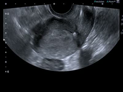 HD for signed-in usersTransvaginal pelvis
IUD Displaced in the Cervical Canal: Ultrasound Findings and Clinical Management
Uterus in anteversion, presenting regular contours and homogeneous myometrial echotexture. Thickened and homogeneous endometrial echo. Note the presence of a metallic intrauterine device (IUD) (compatible with a copper IUD) in an anomalous situation. The upper end of the device is displaced caudally, located at the level of the internal orifice of the uterine cervix (it is interesting that the ultrasound doctor shows here the distance from the upper portion of the IUD to the uterine fundic serosa). There are no textural signs of myometrial perforation or associated intracavitary fluid collections.
Open case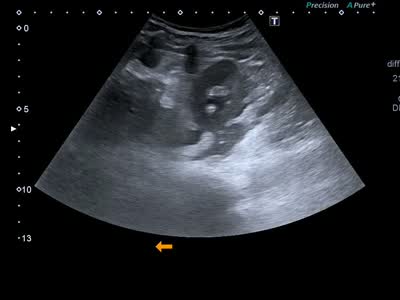 HD for signed-in usersVias Urinárias
Lamina liquida perirrenal e hidronefrose ao ultrassom
Imagem ultrassonografica renal demonstrando a presenca de fina lamina liquida perirrenal, em contexto de sinais de hidronefrose. Nao e evidenciado fator obstrutivo causador na imagem avaliada.
Open case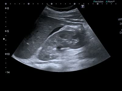 Open case · HDAbdomen / FAST-eFAST
Free fluid in the hepatorenal space and right paracolic gutter
Ultrasound images showing an anechoic collection, consistent with free fluid, in the perihepatic region, in the hepatorenal space (Morison pouch), extending toward the right paracolic gutter.
Open case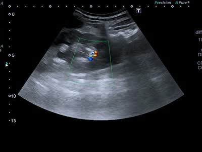 Open case · HDUrinary tract
Non-obstructive nephrolithiasis with twinkling artifact
Daily-practice case showing a non-obstructive stone in the lower third of the left kidney, with a Doppler twinkling artifact.
Open case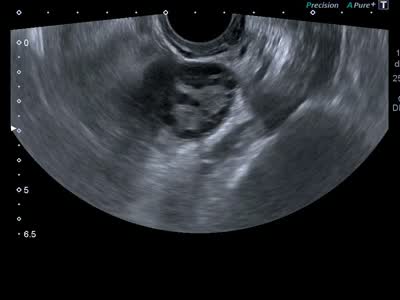 HD for signed-in usersTransvaginal pelvis
Micropolycystic ovary with peripheral follicles
Daily-practice case with an enlarged ovary showing multiple subcentimeter follicles with predominantly peripheral distribution.
Open case